Breeders of cows know that their productivity is influenced by age, breed, overall animal health, nutrition, as well as a number of other factors. Among them - the shape and size of the udder. Experienced breeders have an idea of what the mammary glands should be in order to achieve the greatest amount of milk. Whether the cow will have a high level of milk yield, they are easily determined by the appearance of the glands. We offer you to get acquainted with the structure of the udder, the processes of formation and release of milk.
Udder structure
Udder is the organ of the cow in which milk is produced. There are 2 parts in it - right and left - and 4 mammary glands. The parts are separated by a middle partition. In each of the parts there are 2 lobes — anterior and posterior, which can be developed unevenly. Most often, more milk is formed in the posterior lobes than in the front ones, this is due to the content of more alveoli in them.  Diagram of the udder and secretion section: 1 - deep veins, 2 - deep arteries, 3 - connective skeleton (stroma), 4 - glandular tissue (parenchyma), 5 - superficial saphenous veins and arteries, 6 - milk tank, 7 - nipple tank , 8 - nipple canal opening, 9 - nipple canal, 10 - nipple sphincter, 11 - milk ducts, 12 - bunch of alveoli, 13 - nerves, 14 - myoepithelium, 15 - secretory cells, 16 - duct of the alveoli group.
Udder form 3 types of tissue: glandular, fatty, connective. Glandular tissue is formed by the alveoli. The connective tissue performs a support function, and also protects the udder from the adverse effects of the environment, its fibers divide the milk-forming organ of the cow into lobes.
Each share includes:
- glandular tissue;
- connective tissue;
- milk ducts;
- vessels;
- nerves.
Learn how to properly treat udder swelling in cows.
Circulation
The circulatory system of the udder is represented by:
- perineal arteries;
- external controversial artery and vein;
- vein and artery of the milk tank;
- subcutaneous abdominal milk vein.
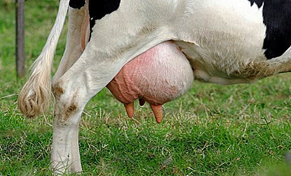
 Through the arteries, the blood enters the mammary gland, through the veins - returns to the heart. Arteries are located deep, they can not be seen, but the veins are clearly visible on the surface of the udder. Powerful subcutaneous abdominal veins, which are well visible, are called milky, and their size determines the milkiness of the cow - the larger they are, the higher the milk yield.
Did you know? In ancient Egypt, cows were not sacrificed, because they were considered the sacred animals of the goddess of heaven and fertility Hathor.
The better the circulatory system in the mammary gland is developed, the more branches it has, the better it is supplied with nutrients and oxygen.
Lymphatic system
The lymph circulation system begins in the area of the alveoli, around which are located lymphatic gaps and spaces. Collection of lymph occurs in interlobular vessels. Later it flows through the lymph nodes into the lymphatic cistern and then through the thoracic duct into the vena cava.  In the mammary glands there are many vessels for lymph flow. Each lobe contains lymph nodes the size of a walnut. The lymph is derived from them by the vessels, one of which is connected with the system of lymphatic circulation of the rectum and genitals, and the other with the inguinal lymph nodes.
Nerves
In the skin, on the nipples, in the alveoli there are many nerve endings that respond to irritation that occurs in the mammary gland, and report them to the brain. The most sensitive nerve receptors are located in the nipples. The spinal cord with an udder is connected by nerve trunks, which branch into thin filaments that conduct signals from the central nervous system. Nerves play an important role in the growth and development of the mammary gland, as well as in the volume of formed milk. 
Milk follicles
Glandular tissue is formed by the alveoli or follicles in the form of tiny sacs. Inside they contain cells in the form of asterisks, responsible for the production of milk. With the help of tubules in which the same stellate cells are located, the alveoli have a connection with the milk ducts. These channels pass into the milk tank, and the tank communicates with the nipple.
Dairy follicles have an extensive working area, a complex system of work. They react sharply to changes in the environment and change every time after lactation. It is in the alveoli before the milking process begins that 50% of the milk accumulates (up to 25 liters). The remaining 50% is contained in the ducts, the milk tank and the nipples.
Read also about how to milk a cow.
Nipples
Each lobe has one nipple. Often, cows can be found 5 and 6 nipples, which can even give a little milk. Udder is considered good if its nipples are of the same size - from 8 to 10 cm long and 2 to 3 cm in diameter, the shape of a cylinder, vertically hang down and perfectly release milk when compressed. The nipple secrete base, body, apex and a cylindrical part. Its walls form the skin, connective tissue, mucous membranes.  At the top is the sphincter, thanks to which the milk does not pour out without milking. Nipples play a major role in lactation and preventing infection in the mammary glands. Their skin does not have sweat and sebaceous glands, so care should be taken to it to avoid reproduction of pathogenic microflora and the formation of cracks.
Important! Shares do not have a message among themselves. Therefore, it is important for the livestock breeder to empty each of them to the end, because the milk cannot move from one lobe to another and leave the other nipple, which means it will not be formed in the maximum amount next time.
Stages of udder development in cows
For the development of the mammary glands of the cow are responsible nervous and endocrine systems. The embryo glands are laid out of the epithelial thickening, located in the abdominal cavity behind the navel. Subsequently, 4-6 hillocks are formed from it, from which, after the formation of the circulatory system and nerve fibers is completed, the mammary glands develop. The udder of a 6-month fetus already has milk ducts, a cistern, a nipple and adipose tissue.  After birth and before puberty, the udder gradually takes shape and grows. During this period, it is mainly formed from adipose tissue. When a cow comes to puberty, her udder increases significantly, which is affected by the active production of sex hormones, and takes the form that is characteristic of a mature chick. The growth of canals and ducts ends by the 5th month of pregnancy, by 6-7 months the alveoli are finally formed.
Glandular tissue is fully formed by the 7th month of pregnancy, its increase will occur after calving. This process will be affected by the active production of hormones, proper milking, massage and nutrition of the heifer. The development and growth of glands is carried out up to 4-6 genera. Changes occur in the structure in accordance with sexual cycles, lactation periods, exercise, and age of the cow.
Important! It is believed that cows with a wide cup-shaped udder, which is well projected forward, adjacent to the body, highly attached at the rear, have high performance. Udder fractions should be even and symmetrical. When palpating, the udder should be soft and supple.
Extinction of the mammary glands occurs after 7-8 births - during this period the volume of glandular tissue and ducts are reduced, and the connective and adipose tissues increase. Successful breeders with proper efforts, which include enhanced nutrition and quality care, can extend the productive period of the heifer to 13-16 lactations, and sometimes even longer. 
How does the process of milk formation
The main function of the udder is lactation. The lactation process consists of two stages:
- Milk formation.
- Milk yield.
Check out the best breeds of dairy cows.
The process of milk formation is influenced by several factors:
- active replenishment of the udder with nutrients through the blood vessels;
- normal functioning of the lymphatic system;
- release of the hormone prolactin as a result of calving, irritation of the nipples when sucking a calf or when warmly touched.
 However, if the udder is not emptied for longer than 12-14 hours, the pressure increases, the action of the alveoli is inhibited, milk production decreases. Thus, with regular and complete emptying of the udder, the level of milk formation is maintained at a high level. Long intervals between milking processes or incomplete emptying of the udder entail a decrease in milk production.
Did you know? The most expensive beef in the world is obtained from Japanese Wagyu cows. The Japanese, who live in the vicinity of the city of Kobe, where these cows were mostly divorced, treated their pets with care - wiped them with sake and drank their beer. As a result, they received very tender and tasty meat, which today is sold at 100 euros for 200 grams of tenderloin.
Milk yield
Milk yield is a reflex that manifests itself during milking and is accompanied by the release of milk from the alveoli into the cisterns. From the milk follicles, the fluid is excreted by compressing the cells that are around them. After such a compression, it flows into the ducts, then into the cistern, the outflow channel and the nipples.
During irritation with the lips of the calf or with other irritable factors of the nipples from their nerve endings, a signal is emitted to the brain of the cow, which gives the command to the pituitary gland. The pituitary gland releases hormones into the bloodstream, which are responsible for the production of milk and the contraction of the myoepithelium of the mammary glands. As a result, there is a reduction of cells located around the alveoli.
The cells, in turn, compress the alveoli, and from them the milk falls along the ducts into the cisterns. Milk production is carried out after 30-60 seconds after irritation of the nipples. Its duration is 4-6 minutes. During this time, the milking process should begin. After the expiration of its oxytocin is no longer produced, the alveoli are not compressed, the reflex milk transfer dies away.  The process of milk delivery is also regulated by some incentives: milking time, voice of a milkmaid, milking machines, etc. Milk output occurs simultaneously in all 4 lobes, even if one nipple is irritated. The smallest amount of milk comes out of the share that is given out last. As a rule, by the time of her milking, the milk flow reflex is already extinct.
Important! It has been empirically established that the greatest milk loss occurs if, when milking a cow, the nipples shrink at a rate of 60-90 times per minute.If a cow is frightened during the lactation, if it is rude to do with it, to cause pain, then the process may stop. In such cases, the ducts are narrowed, and it is possible to milk only the milk contained in the tanks. The milk accumulation process lasts 12-14 hours after the previous milking. The nipple response to irritation occurs after 4 hours. Thus, several factors influence milk yield, the most important of which is a well-developed udder, rich in glandular tissue.
 Milk flow directly affects the development of the circulatory and lymphatic systems. However, not only the udder plays a role in the performance of a cow — a poorly-fed cow, poorly groomed, undernourished, suffering from a deficiency of vitamins and minerals, will not be able to produce enough milk, even with good udder.