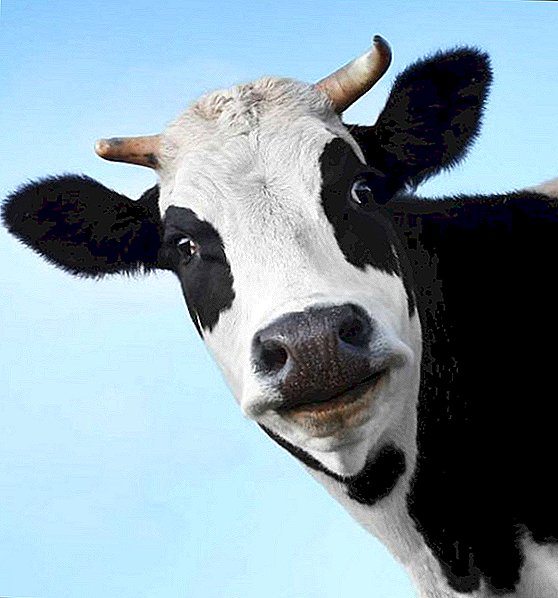
At birth, a cow may experience a situation in which the animal will not be able to do it on its own. In this case, the veterinarian performs the operation - cesarean section. Similar operations are done to people, but the treatment of cattle has its own characteristics.
What is a cesarean section?
A caesarean section is an emergency operation, the purpose of which is to save the life of a cow and help her baby to be born. Its essence is that on the belly of the cows make a cut through which the calf is taken out. This is an efficient and cost-effective operation; It can be carried out not only in clinics, but also in the conditions of an ordinary farm. The percentage of positive outcomes reaches 90%, moreover, it is usually possible to save the lives of both animals.
Important! The consequences of cesarean section do not affect the production of milk and retain the ability for subsequent reproduction of offspring.
Indications for surgery
The decision on surgery is taken by a veterinarian. - after it is established that the cow is not able to give birth in a natural way. Also, indications for surgery are:
- non-disclosure or defective neck opening;
- large fruit weight;
- narrow birth canal;
- twisting of the uterus;
- fetal deformity;
- death of the fetus.
 The most suitable date is 12 hours after the start of the delivery process. The prognosis may worsen if there was injury or infection of the birth canal during the care.
Find out why a cow has a miscarriage, how to properly run a cow in front of calving, and also, read because of what a vagina falls out of a cow.
How to make a cesarean section to a cow
Like any other operation, a cesarean section consists of several successive stages.
Fixing
There are two types of fixation:
 Standing - when the incision is performed on the side of the abdominal wall. The animal is fixed in a special machine, hind limbs are strapped.
- In the prone position - when cutting in the area of the lower abdominal wall. The animal is felled on the operating table (you can use several bales of hay or straw, covering them with a tarpaulin), the hind and fore limbs are strapped with straps, the head is held and pressed to the surface with your hands.
 Standing - when the incision is performed on the side of the abdominal wall. The animal is fixed in a special machine, hind limbs are strapped.
However, it is not uncommon for a standing cow to lie on the ground during an operation.
Preparation of the surgical field
To conduct a high-quality operation, it is necessary to carry out preliminary training, which consists of the following actions:
- Clean hair.
- The area in the incision area is thoroughly washed with soap and then carefully shaved.
- The skin is rubbed to dryness, smeared with alcohol or iodine.
- The incision area is isolated with a clean cloth.
Did you know? In the language of the cow is located 25 thousand taste buds. One individual produces 150 liters of saliva per day and makes about 100 chewing movements.
Antiseptic and anesthesia
For contraction of the uterus and its easier removal from the abdominal cavity, epidural anesthesia is required. The place where the injection is made, is located between the first and next caudal vertebrae. A needle is inserted perpendicular to the skin, and after a puncture, it is moved inward at an angle of 45 °. The correct puncture depth should be about 3 cm. The solution should flow when the syringe is lightly pressed.

Anesthesia can be of different types:
- Low (the back) - used for operations in a standing position. Enter 20 ml of novocaine solution, heated to body temperature.
- High (front) - carried out at the position of the body on the side. Inject 130 ml of anesthetic solution. In this case, paresis of the pelvic limbs occurs.
Operation technique
A caesarean section consists of the following steps:
- Operative access (laparotomy).
- Eventration of the uterus.
- Opening a hole.
- Extraction of the fetus and the separation of the placenta.
- Stitching wounds.
- Closure of the wounds of the abdominal wall.
Incision
Most often, a ventro-lateral incision is performed. It provides good access to the uterus, and at the same time causes a relatively minor injury to the body. It can be done left or right.

The abdominal wall is cut to 35 cm. Start the incision at the level of the nearby edge of the udder 10 cm above its base. The incision is conducted from top to bottom and ends in front of the abdominal wall 4 cm above the main vein of the abdomen, it should be slightly tilted.
After an incision of the skin and fascia, the rectus abdominis is separated along its fibers with the blunt end of a scalpel. Then, in the middle of the wound, take a piece of forceps of the vagina of the straight muscle of the abdomen with forceps and make an incision that coincides with the direction of the skin wound, while opening it and the peritoneum fused with it.
Important! Quickly athide the abdominal cavity or remove peritoneal fluid is strictly prohibited, as the animal may come shock.
Eventration and opening of the uterus
After isolating the wound of the abdominal wall with sterile wipes, the omentum is cut, and only after that the eventus of the uterine horn is transferred. Eventration is called pulling up the horn of the uterus, in which the fetus is located, to the opening. This is done manually - first they grope a limb with a hand, then they seize it with the uterus and pull it on themselves until the tip of the horn comes out of the wound.
Removal of the fetus and the placenta
When all the tissues are cut, the assistant takes hold of the wound edges and pushes them apart, while the veterinarian cuts fetal membranes at this time, releases the amniotic fluid and takes out the baby. If the fetus was in the head presentation, it is removed for the pelvic bones, and if in the pelvic - for the head and chest bones. In a baby, the mouth and nose are cleaned of mucus, and the umbilical cord is also treated. In conclusion, the last place is separated.
Find out why the cow does not depart the last.
Stitching the wound of the uterus and closing the wound of the abdominal wall
 After the fetus is removed together with the afterbirth, you can begin to sew the uterus. This stage is very important, because only if it is done correctly, the further recovery will be easy. After stitching the uterus, the abdominal cavity is examined, tissues are removed and the incision area is thoroughly washed. After the operation is completed, an extrapleural block is performed according to V.V. Mosin or Novocain is administered intravenously.
If it is determined that the fetus was dead during surgery, antibiotics, such as biomitsin or penicillin, are necessarily introduced to avoid the development of peritonitis.
Postoperative care for a cow
After the operation, the animal must be kept separate from others for several days. Antibiotics are injected for 5 days to avoid the risk of inflammation.
The veterinarian conducts an examination after 3 days, checking for signs of postoperative complications.
Did you know? Baby cows and bulls are called calves. However, few people know that the same name in children of bison, bison and even buffaloes.
Thus, a caesarean section is not a very complicated operation that can save a cow and her child. However, it can only be done by a specialist, of course. If necessary, it should be addressed as early as possible, since the main thing is to conduct the operation on time.